Chest Tube Insertion
Urgent drainage procedure for pneumothorax, hemothorax, and pleural effusion management
What is Chest Tube Insertion?
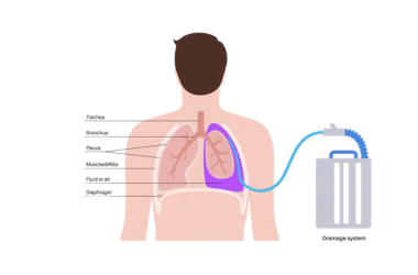
Chest Tube Insertion (Thoracostomy) is an urgent medical procedure where a tube is placed between the ribs into the pleural space to remove air, blood, or fluid. It is often a life-saving procedure performed in emergency situations for conditions like pneumothorax (collapsed lung), hemothorax (blood in pleural space), and large pleural effusions causing respiratory compromise.
The chest tube allows continuous drainage of fluid or air, restoring proper lung expansion and respiratory function. It provides both immediate symptom relief and diagnostic information about the underlying condition requiring the intervention.
Why is Chest Tube Important? It helps:
- Restore lung expansion in pneumothorax
- Drain blood from hemothorax
- Remove infected fluid (empyema)
- Prevent tension pneumothorax complications
- Allow lung re-expansion after drainage
Types of Chest Tubes
Small Bore Tube
14-18 French catheter for air drainage in pneumothorax
Large Bore Tube
28-40 French catheter for blood or thick fluid drainage
Pigtail Catheter
Small flexible tube for pneumothorax and effusion drainage
Needle Aspiration
Immediate decompression for tension pneumothorax
Emergency Tube
Rapid insertion for life-threatening conditions
Indwelling Drain
Long-term drainage for persistent collections
Indications for Chest Tube
Pneumothorax
Collapsed lung requiring air drainage
Hemothorax
Blood collection after trauma
Empyema
Infected pleural fluid drainage
Large Effusion
Symptomatic pleural fluid drainage
Tension Pneumothorax
Life-threatening emergency condition
Post-operative
After thoracic or cardiac surgery
When is Chest Tube Needed?
- Tension pneumothorax (medical emergency)
- Traumatic hemothorax with respiratory compromise
- Large pneumothorax with significant lung collapse
- Infected pleural fluid (empyema) drainage
- Post-surgical fluid or air accumulation
- Persistent spontaneous pneumothorax
- Malignant pleural effusion with symptoms
- Chylothorax drainage
Chest Tube Insertion Procedure
Positioning
Patient positioned supine or lateral position with arm elevated
Anesthesia
Local or general anesthesia administered at insertion site
Insertion
Tube inserted at 4th-6th intercostal space mid-axillary line
Confirmation
Chest X-ray confirms proper tube position and lung re-expansion
What to Expect After Insertion
Management and monitoring after chest tube placement:
- Tube remains in place until drainage resolves
- Regular monitoring of drain output
- Pain management with adequate analgesia
- Strict aseptic technique for dressing changes
- Physical activity restrictions as advised
- Follow-up imaging to assess lung expansion
- Tube removal once criteria met
Note: Chest tube insertion is usually performed in hospital setting under medical supervision. Duration of placement varies based on underlying condition and drainage pattern.
Monitoring After Chest Tube Insertion
Daily Output
Monitor amount, color, and consistency of drainage daily
Vital Signs
Regular monitoring of temperature, blood pressure, and heart rate
Imaging Follow-up
Serial X-rays to assess lung expansion and drainage
Removal Criteria
Tube removed when drainage minimal and lung fully expanded
Why Choose Our Chest Tube Center?
Expert emergency and chest physicians with advanced imaging guidance
Emergency Equipped
24/7 availability for urgent procedures
Experienced Physicians
Specialists in emergency chest procedures
ICU Monitoring
Comprehensive post-procedure care and monitoring
Rapid Response
Immediate intervention for life-threatening conditions
Request a Callback
Get expert advice within 24 hours
Other Diagnostic Tests
Explore our specialized services
Emergency Contact
+91 813-044-8904
24/7 available for respiratory emergencies