EBUS (Endobronchial Ultrasound)
Advanced ultrasound-guided bronchoscopy for lymph node and lung tissue diagnosis
What is EBUS?
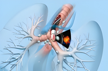
Endobronchial Ultrasound (EBUS) is an advanced diagnostic technique that combines bronchoscopy with ultrasound imaging. A specialized bronchoscope with an integrated ultrasound probe is passed through the mouth to visualize and sample mediastinal (central chest) lymph nodes and lung tissue. This allows for precise, real-time tissue sampling with minimal invasiveness.
EBUS has revolutionized lung cancer staging and diagnosis of lymph node diseases. It provides superior visualization compared to traditional bronchoscopy and significantly reduces the need for more invasive surgical procedures like mediastinoscopy.
Why is EBUS important? It helps physicians:
- Stage lung cancer accurately (assess nodal involvement)
- Diagnose mediastinal lymph node enlargement
- Obtain tissue samples from hard-to-reach areas
- Diagnose sarcoidosis and tuberculosis
- Assess central airway obstruction
Types of EBUS Procedures
Convex-probe EBUS (CP-EBUS)
Used for mediastinal lymph node sampling and lung tissue biopsy.
Radial-probe EBUS (RP-EBUS)
Used for peripheral lung lesion sampling and assessment.
EBUS with TBNA
Transbronchial Needle Aspiration combined with ultrasound guidance.
EBUS-EUS Combination
Combined approach for better access to posterior mediastinal nodes.
Clinical Indications for EBUS
- Suspected lung cancer staging
- Mediastinal lymph node enlargement of unknown cause
- Suspected sarcoidosis
- Tuberculosis diagnosis and confirmation
- Suspected lymphoma or metastatic disease
- Central airway obstruction assessment
- Granulomatous lung disease evaluation
- Immunocompromised patient with lung infiltrates
Preparation for EBUS
Pre-procedure Assessment
Blood tests, chest X-ray, and CT scan review required before procedure.
Fasting
Nil by mouth (NPO) for 6-8 hours before the procedure.
Medication Management
Inform doctor about blood thinners and antiplatelet medications.
Sedation Discussion
Meet anesthesiologist to discuss sedation options and risks.
What to Expect During EBUS
EBUS typically takes 30-60 minutes depending on complexity:
- Moderate sedation or general anesthesia is administered
- Specialized EBUS bronchoscope is advanced through mouth
- Real-time ultrasound visualization of lymph nodes
- Needle aspiration or tissue samples obtained
- Samples sent to pathology for analysis
- Recovery in post-operative area with monitoring
Comfort: Sedation ensures comfort during the procedure. Most patients have minimal memory of the procedure and experience minimal discomfort.
Post-EBUS Care
Recovery Monitoring
Recovery period of 1-2 hours with vital sign monitoring.
Diet Resumption
Clear liquids after 2 hours, normal diet once fully alert.
Activity Restrictions
Rest for 24 hours. No driving or operating machinery for 24 hours.
Pathology Results
Tissue samples analyzed. Results available in 5-7 days typically.
Why Choose Our EBUS Center?
Advanced interventional pulmonology with expert care
Expert Pulmonologists
Specialized in advanced EBUS procedures
Latest Technology
High-definition ultrasound systems
High Diagnostic Yield
Superior sample quality and accuracy
Same-Day Discharge
Minimally invasive with quick recovery
Request a Callback
Get expert advice within 24 hours
Other Diagnostic Tests
Explore our specialized services
Emergency Contact
+91 813-044-8904
24/7 available for emergencies